
Home / Medical Article / Mechanisms by Which Heavy Metals Cause Toxicity in the Body
Why can heavy metals and certain metalloid compounds – such as lead, mercury, cadmium, arsenic, or certain forms of chromium – affect multiple systems throughout the body, from the blood and blood vessels to the kidneys, brain, bones, intestines, and hormonal system?
The answer lies in their mechanisms of toxicity at the cellular level. These substances do not affect only one organ. They can interfere with fundamental processes shared by cells throughout the body, including mineral utilization, enzyme activity, energy production, free-radical regulation, gene expression, and cell-to-cell communication.
When these processes are disrupted, effects can occur across multiple body systems. However, the pattern and severity of toxicity are not the same in everyone. They depend on the substance and its chemical form, the dose and duration of exposure, as well as individual factors and susceptibility.
To understand why these substances can have such widespread effects, let us examine the key mechanisms underlying heavy-metal toxicity.
The body requires minerals such as calcium, zinc, iron, copper, and magnesium as components of many enzymes, receptors, and proteins. Some toxic metals have electrical charges or chemical properties similar to these essential minerals. As a result, they may use the same transport pathways or occupy binding sites normally reserved for essential minerals. This mechanism is known as molecular and ionic mimicry.
This can be compared to a counterfeit key that closely resembles the real one. It may be able to open the door and enter the cell, but once inside, it can cause the cellular machinery to malfunction. Enzymes may work more slowly, energy production may decline, or antioxidant defenses may become less effective.
For example, zinc supports the activity of more than 300 enzymes in the body. One illustrative example is alcohol dehydrogenase, an enzyme involved in alcohol metabolism. Zinc must bind to this enzyme to accelerate the reaction involved in alcohol breakdown. Under conditions in which heavy metals have accumulated in the body, heavy metals may compete for the binding sites normally occupied by zinc. If a heavy metal binds to the enzyme in place of zinc, the enzyme may no longer function properly, potentially interfering with the process of alcohol metabolism.

Cells use oxygen and nutrients to generate energy, producing free-radical molecules as by-products. Under normal conditions, the body can regulate and eliminate free radicals. When toxic heavy metals accumulate, however, they may increase free-radical generation to the point that free radicals exceed the body’s capacity to control them and begin damaging cells. This condition is known as oxidative stress.
Iron, copper, certain forms of chromium, vanadium, and cobalt can generate free radicals directly, including through Fenton or Fenton-like reactions. In such reactions, antioxidants such as vitamin C may themselves participate in pro-oxidant chemistry under particular conditions.
Lead, cadmium, and mercury can increase free radicals indirectly through several important mechanisms:
They can therefore be compared to both “adding sparks” and “reducing the effectiveness of the fire extinguisher” at the same time. When free radicals exceed the body’s ability to control them, downstream effects may include:

Many proteins contain thiol or sulfhydryl groups (-SH), which play important roles in protein structure, folding, and function. Certain metals, particularly mercury, cadmium, and some forms of arsenic, can bind to these sites.
When critical sites on proteins are occupied, the proteins may change shape or fail to function normally. The consequences can involve enzymes, antioxidant systems, cell-surface receptors, transport proteins, and hormone-signaling pathways. Important examples include peptide hormones such as insulin and growth hormone. Some heavy metals may interfere with insulin signaling, contributing to insulin resistance and diabetes-related mechanisms, while some may alter the structure or function of growth-hormone-related proteins.
Lead does more than make red blood cells more fragile. It also inhibits key enzymes in heme synthesis, including delta-aminolevulinic acid dehydratase (ALAD) and ferrochelatase. This reduces hemoglobin production and can shorten red-blood-cell lifespan. At high levels of lead exposure, increased red-cell destruction may also occur.
Many proteins contain thiol or sulfhydryl groups (-SH), which play important roles in protein structure, folding, and function. Certain metals, particularly mercury, cadmium, and some forms of arsenic, can bind to these sites.
When critical sites on proteins are occupied, the proteins may change shape or fail to function normally. The consequences can involve enzymes, antioxidant systems, cell-surface receptors, transport proteins, and hormone-signaling pathways. Important examples include peptide hormones such as insulin and growth hormone. Some heavy metals may interfere with insulin signaling, contributing to insulin resistance and diabetes-related mechanisms, while some may alter the structure or function of growth-hormone-related proteins.
Lead does more than make red blood cells more fragile. It also inhibits key enzymes in heme synthesis, including delta-aminolevulinic acid dehydratase (ALAD) and ferrochelatase. This reduces hemoglobin production and can shorten red-blood-cell lifespan. At high levels of lead exposure, increased red-cell destruction may also occur.
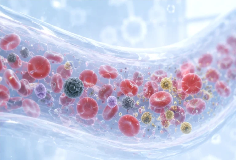
The inner lining of blood vessels, or endothelial cells, produces nitric oxide (NO), which helps blood vessels relax, reduces platelet adhesion, and regulates inflammation.
Toxic metals may increase reactive oxygen species (ROS), which can react with NO and reduce its bioavailability. They may also interfere with endothelial nitric oxide synthase (eNOS). The result can be endothelial dysfunction, including:
However, toxic metals are only one of many factors that may increase risk. Detecting heavy metals in the body does not mean that every individual will develop heart disease or stroke. The source text notes preliminary evidence suggesting that reducing heavy-metal burden may be associated with a lower incidence of coronary and cerebrovascular events.
Lead can accumulate within the mineral structure of bone and remain there for long periods. Bone therefore acts like an internal “storage depot” for lead.
When bone resorption increases – for example during pregnancy, breastfeeding, estrogen deficiency, or osteoporosis – some stored lead may be released back into the bloodstream along with calcium. Internal exposure may therefore occur even after environmental exposure has decreased.
Toxic metals may also affect bone through other mechanisms, such as:
Meta-analyses have reported associations between cadmium and lead exposure and the risk of low bone density or osteoporosis. However, study findings are highly variable and do not establish metals as the sole causal factor.
Different metals cross the placenta to different degrees. Lead and methylmercury can pass from mother to fetus relatively readily, whereas the placenta can retain some cadmium but does not provide complete protection. In summary, several toxic metals can cross the placenta or impair placental function, with the effectiveness of the placental barrier varying by metal. Lead and mercury are important concerns because of their potential for fetal exposure.

The gut microbiota plays roles in digestion, metabolite production, intestinal-barrier integrity, and immune regulation. Heavy metals may alter the composition and function of these microorganisms, resulting in gut dysbiosis. They may also weaken the intestinal barrier and contribute to increased intestinal permeability, often referred to as “leaky gut.”
However, human evidence remains limited and study findings are inconsistent. The source text cites a recent systematic review that included 12 human studies involving arsenic, lead, mercury, and cadmium, and describes possible links between some heavy metals and allergic conditions, chronic fatigue syndrome, and dementia.
Some metals can genuinely trigger allergic reactions. Nickel, cobalt, and chromium ions can act in a hapten-like manner by binding to or modifying the body’s proteins so that the immune system recognizes them as foreign, potentially causing allergic contact dermatitis.
Some substances can act as endocrine disruptors. Toxic metals may interfere with hormonal systems at several points, including:
Cadmium is one example that has been studied as a metalloestrogen because it can activate estrogen-receptor signaling in certain animal and cell models. However, human data remain limited.
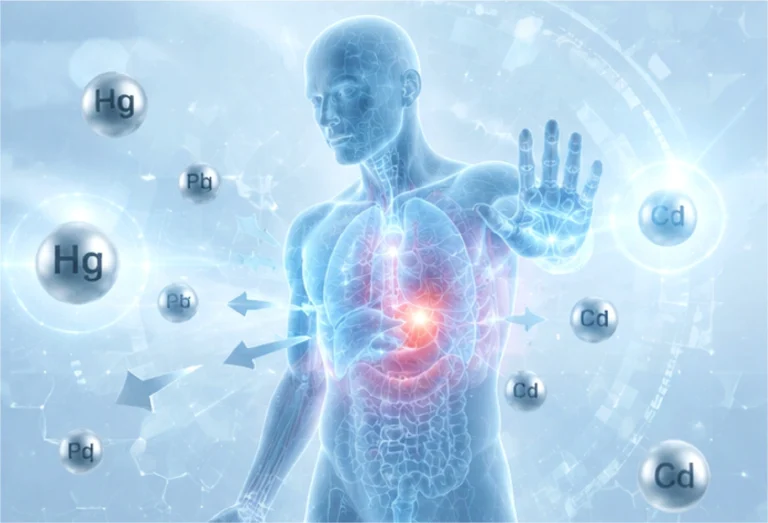
Based on the evidence summarized in the source document, different chemical forms of heavy metals may affect different target organs, as outlined below.
Metal / Chemical Form | Major Target Organ(s) | Key Damage / Effect | Evidence Level |
Lead (Pb) | Brain and nervous system, especially the developing CNS | Neurodevelopment, cognition, synaptic/Ca2+ signaling | A |
Lead (Pb) | Hematologic system | Inhibition of heme synthesis; anemia at high exposure | A |
Lead (Pb) | Kidneys | Chronic tubulointerstitial injury / renal dysfunction | A |
Lead (Pb) | Cardiovascular system | Hypertension, endothelial dysfunction | A-B |
Lead (Pb) | Bone | Long-term reservoir of Pb | A |
Lead (Pb) | Reproductive system | Fertility and reproductive toxicity | B |
Lead (Pb) | Stomach | Gastric cancer | B |
Elemental mercury (Hg0 vapor) | Brain | Tremor; neurobehavioral/neuropsychiatric toxicity | A |
Elemental mercury (Hg0 vapor) | Kidneys | Renal accumulation and nephrotoxicity | A |
Inorganic Hg2+ | Kidneys, especially renal tubules | Nephrotoxicity | A |
Methylmercury (MeHg) | Brain and CNS | Sensory, motor, and cognitive neurotoxicity | A |
Methylmercury (MeHg) | Developing fetal brain | Neurodevelopmental toxicity | A |
Cadmium (Cd) | Proximal renal tubules | Tubular proteinuria -> Fanconi-type dysfunction in severe exposure | A |
Cadmium (Cd) | Bone | Osteomalacia / osteoporosis and bone fragility | A |
Cadmium (Cd) | Lungs when inhaled; lung cancer | Pneumonitis/chronic pulmonary toxicity; occupational carcinogenic risk | A |
Cadmium (Cd) | Liver | More prominent in acute/high-dose toxicity and accumulation | B |
Cadmium (Cd) | Cardiovascular system | Vascular/CV associations | B |
Cadmium (Cd) | Pancreas, stomach, prostate | Cancer associations | B |
Inorganic arsenic (As) | Skin | Hyperpigmentation, hyperkeratosis, and skin lesions | A |
Inorganic arsenic (As) | Peripheral nervous system | Sensorimotor neuropathy | A |
Inorganic arsenic (As) | Cardiovascular system | Vascular disease / cardiovascular risk | A-B |
Inorganic arsenic (As) | Skin, lung, bladder | Established human carcinogenic targets | A |
Inorganic arsenic (As) | Liver | Hepatic injury / portal abnormalities in some exposure settings | B |
Inorganic arsenic (As) | Kidneys | Renal effects | B |
Inorganic arsenic (As) | Metabolic/endocrine system | Association with diabetes/metabolic dysfunction | B |
Inorganic arsenic (As) | Lung, bladder, non-melanoma skin | Cancer | A |
Inorganic arsenic (As) | Kidney, prostate | Cancer associations | B |
Hexavalent chromium Cr(VI) | Respiratory tract / lung | Airway injury, inflammation, fibrosis, and lung carcinogenesis | A |
Hexavalent chromium Cr(VI) | Nose and sinuses | Irritation and cancer risk | A |
Hexavalent chromium Cr(VI) | Skin | Ulceration/contact injury | A |
Hexavalent chromium Cr(VI) | Kidneys and liver | Organ injury | B |
Nickel compounds | Skin | Allergic contact dermatitis | A |
Nickel compounds | Lungs and respiratory tract | Inflammation/fibrosis in occupational exposure | A |
Nickel compounds | Lung and nasal cavity | Risk particularly associated with nickel refining and certain nickel compounds; should not be generalized to all metallic nickel | A |
Excess manganese | Basal ganglia of the brain | Manganism; extrapyramidal motor dysfunction | A |
Excess cobalt | Heart | Cardiomyopathy in high systemic exposure | A-B |
Excess cobalt | Thyroid | Hypothyroid effects | A-B |
Excess cobalt | Hematologic system | Polycythemia from erythropoietic effects | A |
Excess cobalt | Lungs | Interstitial lung disease, especially with tungsten-carbide co-exposure | A |
Excess cobalt | Hearing, vision, peripheral sensory/motor nerves | Toxicity reported in severe systemic exposure or metal-on-metal implants | B |
Aluminum overload | Bone | Osteomalacia/adynamic bone disease, especially in renal failure/dialysis exposure | A |
Aluminum overload | Central nervous system | Dialysis encephalopathy/high systemic exposure | A |
Aluminum overload | Bone marrow | Microcytic anemia in significant systemic accumulation | A-B |
Aluminum overload | Breast | Mammary epithelial oxidative stress, genomic instability, estrogenic signaling | C, predominantly preclinical |
Thallium (Tl) | Peripheral nervous system | Painful sensory-motor neuropathy | A |
Thallium (Tl) | Gastrointestinal tract | Abdominal pain/gastroenteritis in acute poisoning | A |
Thallium (Tl) | Hair and skin | Characteristic delayed alopecia | A |
Thallium (Tl) | Brain, heart, kidneys | Severe multisystem poisoning | B |

Heavy metals are naturally occurring substances that can be present in soil and may enter plants, water supplies, and the air. After entering the body, some can accumulate gradually. Early exposure may produce no obvious symptoms, while continued or substantial exposure may damage cells and can produce manifestations that overlap with those of chronic diseases.
The source document states that when illness is related to a high accumulated heavy-metal burden, reducing that burden may improve symptoms. It also states that the World Health Organization has compiled evidence concerning heavy-metal toxicity and that there may be no safe level for certain toxic metals. The document further notes that evidence regarding the clinical benefits of heavy-metal detoxification remains limited because systematic studies are lacking, while preliminary evidence has suggested improvements in some conditions – including allergic or hypersensitivity symptoms, kidney disease, cardiovascular disease, and neurodegenerative conditions – when elevated heavy-metal burden is demonstrated at baseline.